BrainGFM: AI Model Revolutionizes Brain Imaging with Generative Foundation
Summarized by AI from reporting by ArXiv cs.AI, published under our editorial policy.
Researchers developed BrainGFM, an AI model that improves brain imaging tasks by generating synthetic data and adapting to different imaging conditions. It showed significant improvements in stroke diagnosis and could transform clinical brain imaging.
Key takeaways
- BrainGFM is a generative foundation model trained on 1.3 million MRI and CT slices to improve brain imaging tasks.
- The model demonstrated a 14% relative improvement in radiologist stroke-subtyping accuracy in a human-AI collaboration study.
- BrainGFM-generated FLAIR images approached the diagnostic quality of acquired FLAIR images in blinded reader assessments.
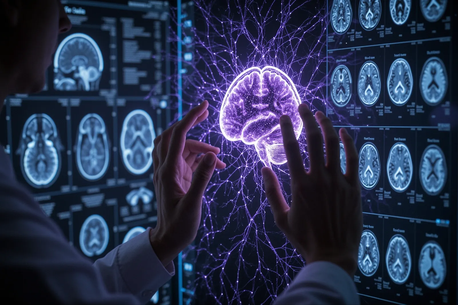
Researchers introduced BrainGFM, a generative foundation model designed to enhance computational brain imaging. This AI model leverages a corpus of 1.3 million MRI and CT slices to improve task-aligned data synthesis and image reconstruction, addressing the scarcity and heterogeneity of clinical brain imaging data.
How BrainGFM Works
BrainGFM extends knowledge transfer beyond traditional representation reuse by utilizing a reusable and reconfigurable brain-imaging prior. This approach allows the model to generate synthetic data for low-data predictive learning and adapt to various imaging conditions through task-adapted post-training. The model was tested across 48 downstream tasks, demonstrating improved segmentation and classification under limited supervision and high-fidelity image enhancement.
Traditional brain imaging models often rely on fixed architectures and entangled features, which limit their adaptability to diverse tasks. BrainGFM's generative approach overcomes these limitations by creating a flexible prior that can be reconfigured for specific tasks. This makes it particularly useful in clinical settings where data is often scarce and varied.
Clinical Validation and Performance
The model underwent rigorous clinical validation involving 4,796 stroke patients across two internal and four external centers. Blinded reader assessments showed that BrainGFM-generated FLAIR images approached the diagnostic quality of acquired FLAIR images. In a human-AI collaboration study, BrainGFM assistance led to a 14% relative improvement in radiologist stroke-subtyping accuracy. These results highlight the model's potential to enhance diagnostic accuracy and efficiency in clinical settings.
FLAIR (Fluid-Attenuated Inversion Recovery) imaging is a crucial tool in diagnosing stroke and other brain conditions. The ability of BrainGFM to generate high-quality FLAIR images that are nearly indistinguishable from acquired images is a significant advancement. This capability can reduce the need for repeated imaging sessions, thereby lowering costs and patient discomfort.
Why It Matters for Everyday People
For patients and healthcare providers, BrainGFM represents a significant advancement in brain imaging technology. By improving the quality and availability of diagnostic images, it can lead to earlier and more accurate diagnoses, particularly for conditions like stroke. This could result in better treatment outcomes and reduced healthcare costs. For radiologists, the model offers a powerful tool to enhance their diagnostic capabilities and reduce the workload associated with interpreting complex imaging data.
Stroke is a leading cause of disability and death worldwide. Early and accurate diagnosis is crucial for effective treatment. BrainGFM's ability to improve stroke-subtyping accuracy can help clinicians make more informed decisions, potentially saving lives and improving patient outcomes. Additionally, the model's adaptability to various imaging conditions makes it a versatile tool for a wide range of neurological conditions.
What You Can Do Today
While BrainGFM is primarily a research tool at this stage, you can stay informed about advancements in AI-driven medical imaging by following research publications from the authors' institutions. If you are a healthcare professional, consider exploring AI-assisted diagnostic tools that may become available in the future. For the general public, understanding the potential of AI in medical imaging can help you appreciate the evolving landscape of healthcare technology.
If you are interested in the technical details, you can access the full research paper on arXiv. For those in the medical field, discussing the potential applications of BrainGFM with colleagues and staying updated on the latest research can provide valuable insights into the future of medical imaging.
Frequently asked
- Is BrainGFM currently available for clinical use?
- No, BrainGFM is currently a research tool and not yet available for clinical use.
- How does BrainGFM improve diagnostic accuracy?
- BrainGFM improves diagnostic accuracy by generating high-fidelity synthetic data and adapting to various imaging conditions, which enhances segmentation and classification tasks.
- Can BrainGFM be used for conditions other than stroke?
- The paper focuses on stroke diagnosis, but the model's adaptability suggests potential applications for other brain imaging tasks.